The Precursor of Breast Cancer
The Precursor of Breast Cancer
DUCTAL CARCINOMA IN SITU
Breast cancer is the leading cancer affecting women in South Africa, and the world. The latest CANSA statistics revealed that 1 in every 26 women are at risk of being diagnosed with breast cancer in South Africa.

What is Ductal Carcinoma in situ?
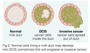
Ductal Carcinoma in situ (DCIS) is a non-invasive type of breast cancer which starts within the milk ducts. It is the earliest form of breast carcinoma – Stage 0. Non-invasive means that abnormal cells are present, but it is still confined to the milk ducts. DCIS has increased risk of developing into an invasive ductal carcinoma (IDC) if left untreated. IDC is the most common type of breast cancer.
Not all DCIS lesions will develop into IDC, it is however unpredictable. DCIS accounts for up to 25% of newly diagnosed breast carcinoma. Overall, DCIS has an excellent prognosis when diagnosed and treated early.
Causes
The exact cause of DCIS has not yet been established. DCIS forms when genetic mutations occur in the DNA of breast duct cells. The genetic mutations cause the cells to appear abnormal, but the cells do not have the ability to break out of the breast duct yet.
Researchers are still working on determining the exact trigger that causes the development of these abnormal cells that eventually lead to DCIS. Your lifestyle, environment and genetic make-up are all thought to play a role.
Risk factors for developing DCIS
- Increasing age – As your age increases, so will your risk of being diagnosed with DCIS (generally uncommon before the age of 30)
- Race – DCIS is less common among African, American, Asian and Hispanic women
- Positive family history will increase your risk for DCIS
- Personal history of benign breast disease, such as atypical hyperplasia
- Parity – There is an increased risk if you did not have any children. Increased risk has also been linked to having children after the age of 30.
- Menarche (the start of menstruation) before the age of 11
- Onset of menopause after the age of 55
- Previous DCIS
Statistics
At Keystone Radiology, we currently diagnose about 1 DCIS patient every month. These lesions were only detected because the patients came for yearly mammograms, enabling us to notice even the smallest changes when comparing their current mammogram to previous imaging. DCIS lesions as small as 0.3 – 0.4 cm have been diagnosed. Most of the suspicious lesions detected were removed completely with our brand-new technology, Vacuum Assisted Breast Biopsy. The main goal is to detect and remove DCIS as soon as possible, thereby preventing the development of invasive breast carcinoma.
How we detect it
DCIS is rarely detected with breast self-examination. Statistics show that only 13% of DCIS presents symptomatically. Annual Mammography and Ultrasound screening gives patients the ease of mind that your breasts are clear of any lesions and breast carcinoma or the forerunner thereof (DCIS). This however, does not underplay the role of monthly breast self-examination.
How we diagnose it
The exact nature of a lesion can only be determined with a screening modality. Whether ultrasound, mammography or both methods will be used is patient specific. Thereafter, suspicious lesions can further be investigated with a biopsy.
What is a breast biopsy
The area of interest is injected with local anaesthetics. A needle is then inserted to remove several samples of tissue from the area that looks suspicious. The samples are sent to a pathologist to obtain an accurate diagnosis. According to the stage of DCIS/cancer diagnosed, an appropriate treatment route will be established.
How it is treated
The treatment plan differs depending on the stage of the disease.
There are three grades of DCIS:
- low or grade I –
- moderate or grade II
- high or grade III.
The lower the grade, the more closely the cancer cells resemble normal breast cells and the more slowly they grow.
For many years, Mastectomy has been the gold standard. With research and more information on breast cancer, a mastectomy is rarely necessary and DCIS and even some forms of breast cancer are treated much more conservatively. Complete removal of the lesion (lumpectomy) represents acceptable treatment for selected patients. In other patients, complete removal of the lesion and hormonal treatment is necessary.
What you can look out for
Immediate mammographic and ultrasound evaluation is indicated should one of the following symptoms present:
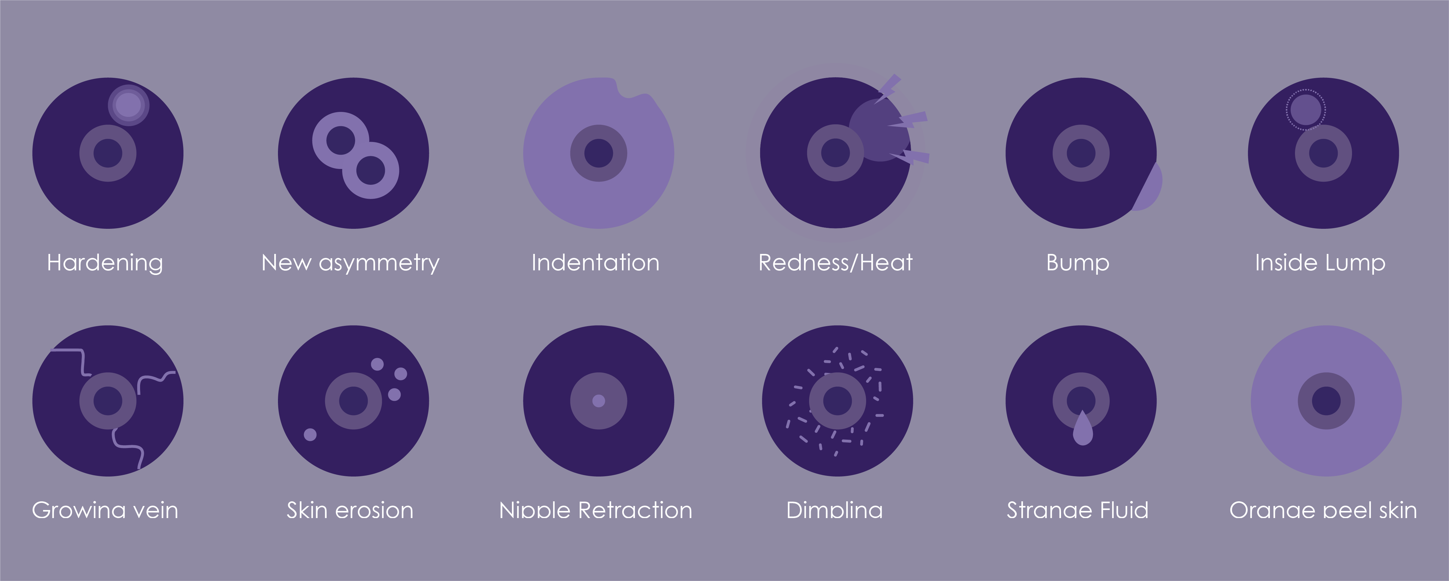
When you are examining your breasts, it is essential to identify the following signs:
- New Lumps.
- Swelling (inflammation).
- Skin changes, especially centrally.
- Breast or nipple pain.
- Redness, scaling or thickening of the areolar area.
- Any discharge
- Unusual swelling under your armpit.
- New size changes.
- Development of new asymmetry.
DCIS is not a diagnosis to fear, as it is treatable and curable. Our mandate at Keystone Radiology is not only to detect breast carcinoma, but to diagnose the precursors of cancer. Annual screening with mammography and ultrasound allows us to ensure your breasts are clear of any abnormalities. Should there be anything to be concerned about, the sooner it is diagnosed, the sooner we can treat and manage it for the absolute best prognosis.
Make a booking
Need to get a Mammogram done?
For further reading, feel free to follow the links below: